Aneurysm Detection | CVisionLab


Aneurysm Detection

Screening for Brain Aneurysms

Screening for Brain Aneurysms


The current state of healthcare is still experiencing difficulties with ineffective diagnostic testing and treatment. The key to mitigating this issue is to discover diseases in their earliest stages.

The pioneering screening technology we are working on has the potential to fulfill this demand. By catching specific conditions based on medical imagery, the technology will be able to guide physicians’ decisions and improve success rates of treatments.


Technological Advancements and Their Impact on the Quality of Healthcare

Correct diagnosis largely depends on the quality of medical image processing. Over the last several decades, images from X-ray, MRI, CT scans have become a viable, sometimes the primary source of diagnostic information. While the performance of human operators is sufficient enough, efficiency can be enhanced by computer vision mechanisms.

Treatment options become significantly more effective when the symptoms are recognized early.
In some cases, it is critical to the patient's wellbeing. However, some changes may not be visible to the human eye, or the image is not processed quickly enough.

The sooner the problem is diagnosed, the better – it is easier to treat, leads to better success rates, reduces the costs, etc.

Here is an example of how timely detection can contribute to a better outcome.

Computer Vision for Aneurysms detection

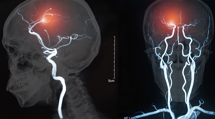
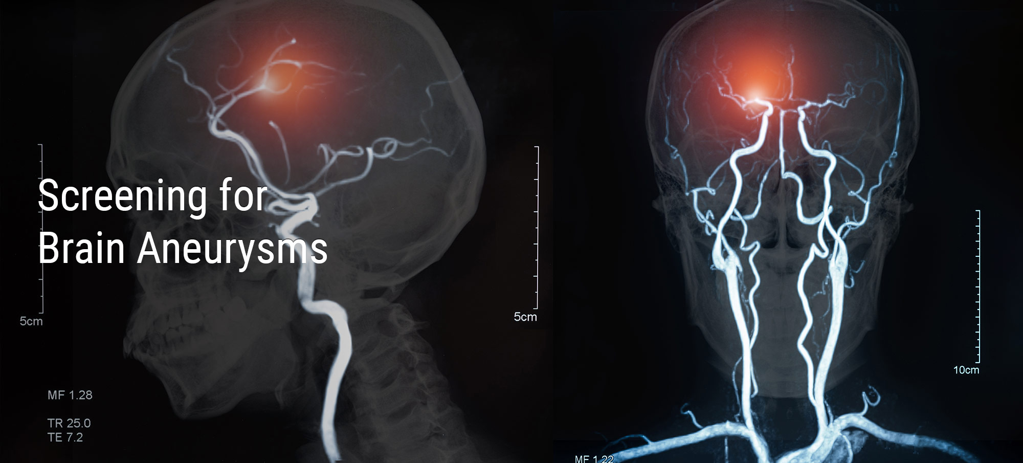
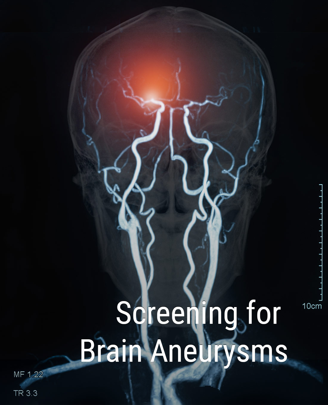
One-third of cases of brain hemorrhage results in death. However, one of its precursors is aneurysm – bulging or enlargement of the artery in the brain. This deformation can be determined by the MRI/CT scanning results.

Unfortunately, the processing of scans can take up some time and worsen the situation.

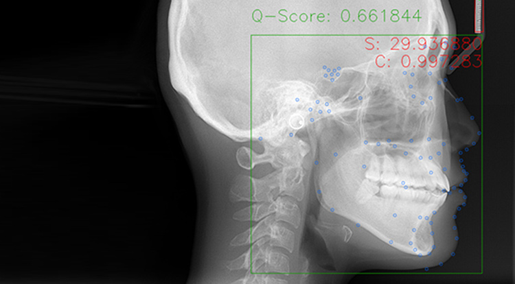
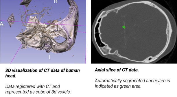
The goal of our project is to identify potentially problematic areas automatically. It will allow medical specialists to have pre-examined imagery processed by high-accuracy detection mechanisms. Thus, they will be able to proceed to treatment with fewer risks of mistakes.


Technical Overview of Our Solution


0.84

Jaccard index

To translate the insight from the example above into a reliable assessment tool, we utilized network segmentation. As part of a trial version, the network was taught based on 300 scans. After receiving an input scan, it segmented the image into two subnetworks – a healthy and sick voxel (an equivalent to pixel but in three-dimensional space). We used the Jaccard index to gauge similarity in sample sets and achieved 0.84. To put it into perspective, the measurement above 0.73 is considered good, and 0.92 is considered excellent.

In the case of aneurysm detection, it is essential to segment any suspicion of the disease. As soon as the symptoms are spotted, the results are transferred to a doctor. The produced segmentation of sick voxels helps them make a timely decision. To increase the precision of the network, we reinforced penalties for false positives.


Challenges of Computer Vision-Assisted Diagnosis

When introducing CV systems into the medical field, we face the underlying problems associated with all automated and machine learning mechanisms. The limitations include the labor-consuming nature of collecting data, ethical ramifications, and sociocultural barriers.

Peer review quality assurance serves as a vital arbiter in any clinical evaluation. Before applying the technology directly into the clinical environment, developers should generate robust evidence and gather massive amounts of data. Also, the tech should be understandable enough so that healthcare providers will be able to operate it correctly.

Future solutions should preserve vigilance about risks and potential misdiagnoses. The challenge is to maintain the same standards during development as well as after getting on the market. And after rolling out new algorithms, the solutions will need to be re-evaluated.


Outcomes and Impacts of CV Detection

By now, there is limited artificial intelligence and computer vision application on the market. Notably, there is no full automation of the medical process. But in reality, it is not the main goal of current CV advancements. The process of collecting patient data and expanding the body of reference material is the main focus and driving force of future solutions.

The technology is far from being mainstream and is yet to be used in real clinics. But despite being at the prototype stage, the current results are already promising. The following years will show how machine learning systems will unravel.

What is already known is that the technology is not aiming to replace human doctors. The main objective will still be assistance, medical image processing and accurate detection of symptoms. With the help of automation, healthcare providers will be able to devote more time to important tasks and leave aside repetitive work.


GET IN TOUCH

By clicking "SEND", I consent to the processing of my personal data.
For more info see our privacy policy.


CVisionLab

Copyright © 2024. CVisionLab LLC
Terms of UsePrivacy Policy
Design-studio cCube.ru. Website development and support